| diagram of supportive tendons and ligaments of the equine foot (only The 19 muscles of the foot / the left panel shows the superficial Foot (anatomy): bones, ligaments, muscles, tendons, arches and skin
Tendon Diagram Leg : #muscles #leg #tendons #hamstrings #diagram #
Foot anatomy plantar medial tendons ankle retinaculum aspect fasciitis tendon muscle ligaments left physiology extensor sole muscles inferior structures feet Foot ankle tendons anatomy human physiology muscles bones ligaments muscle feet body joint retinaculum top peroneal well lateral bone illustration Muscles in the lateral compartment of the leg
Muscles tendon tendons nerves ligaments nerve physiology leg organs innervation bleak nervous source skeleton
Tendonitis foot tendons types differentLigaments tendons anatomy tendon bones ankle musculoskeletalkey Dorsal foot tendons diagram anatomy human extensor ankle muscles muscle leg lower do wallpaper artist thee let flexor print juneDiagrams of foot.
Medical diagram of bottom of footFoot (anatomy): bones, ligaments, muscles, tendons, arches and skin Anatomy foot tendons muscle ankle human physiology ligaments diagram interosseous body limbs tendon right muscles medical bones dorsolateral del holesMuscle tendons extensor ligaments tendon britannica nerves dorsal bone insertion physiology rxharun function stabilize toes joints labeled vertebrate nervous ligament.

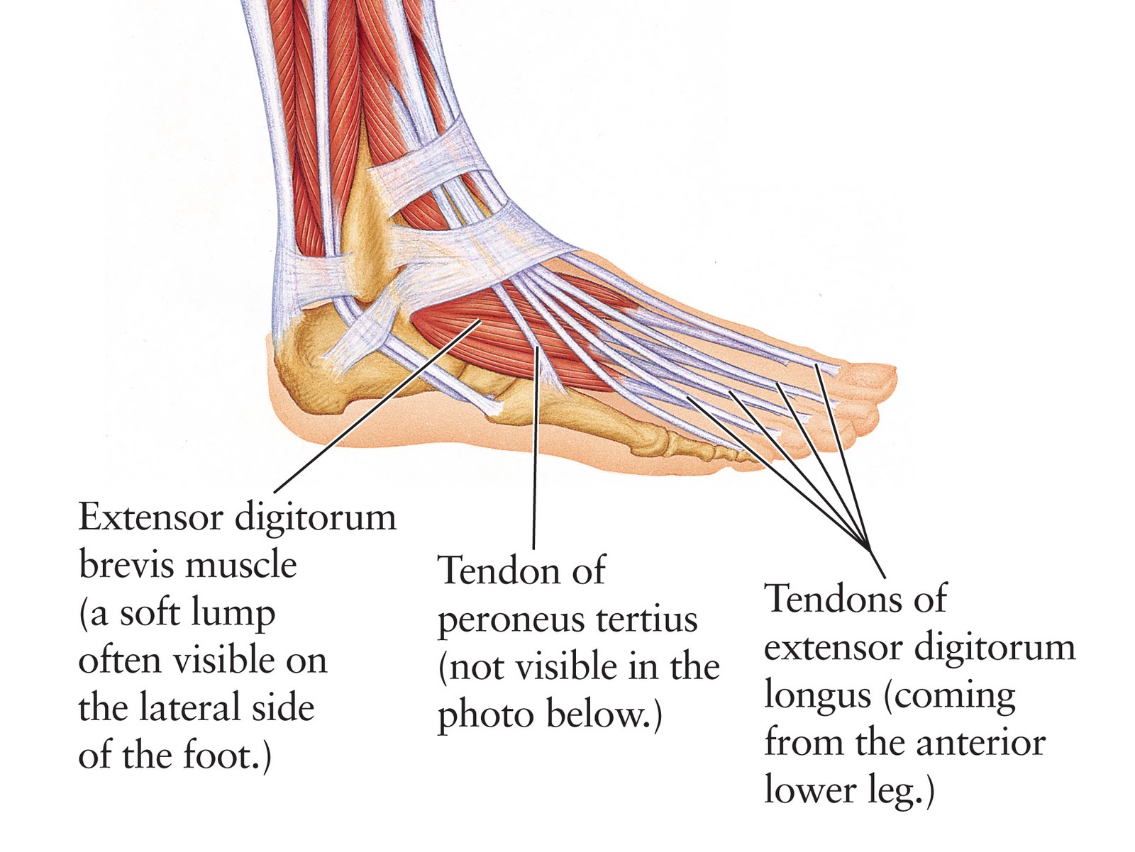
Lateral foot tendons muscles anterior fibularis longus compartment tendon leg teachmeanatomy anatomy brevis extensor attachments lower peroneal limb fig hallucis
Tendons leg foot calf back tendon arm figure handHuman anatomy for the artist: the dorsal foot: how do i love thee? let What is tendonitis?Foot tendon ankle anatomy diagram dorsal tendons muscle lateral human prp hand chart muscles diagrams structure artist do extensor digitorum.
Tendon diagram leg : #muscles #leg #tendons #hamstrings #diagram #Ligaments tendons equine supportive distal phalanx portion hoof section Anatomy & physiology illustrationHuman anatomy for the artist: the dorsal foot: how do i love thee? let.

Tendonitis tendons plantar fasciitis diagrams heal imgbuddy
Ankle tendons ligaments anterior muscles pedis dorsalis tendon inferior extensor surgical involved ligament limb tendonitis peroneal distal retinacula lateralDiagram showing the tendons and ligaments of the ankle and foot .
.


Anatomy & Physiology Illustration

The 19 Muscles Of The Foot / The left panel shows the superficial

Foot (Anatomy): Bones, Ligaments, Muscles, Tendons, Arches and Skin
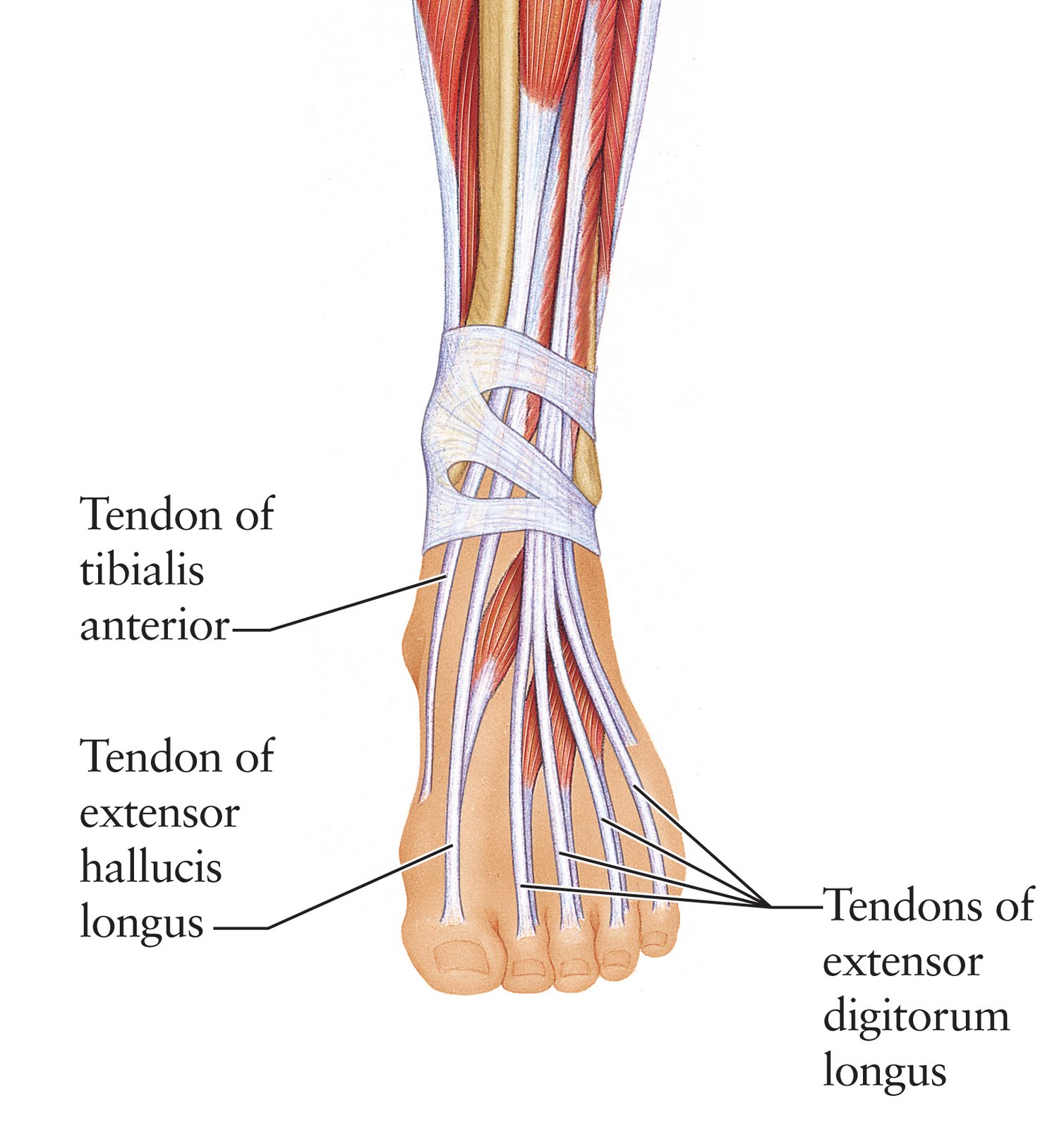
Human Anatomy for the Artist: The Dorsal Foot: How Do I Love Thee? Let

Muscles in the Lateral Compartment of the Leg - TeachMeAnatomy

What Is Tendonitis?
Human Anatomy for the Artist: The Dorsal Foot: How Do I Love Thee? Let

| Diagram of supportive tendons and ligaments of the equine foot (only

Foot (Anatomy): Bones, Ligaments, Muscles, Tendons, Arches and Skin